
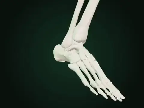
Type II accessory navicular
About this resource
A separate illustrative ossicle sits beside the medial navicular tuberosity. The gap represents an unmodeled synchondrosis. Shown on the moving joint. Illustrative shape only: playback, contact, pressure and symptoms are not changed or calculated.
Compare the illustrated bone shape with typical atlas anatomy as the joint moves.
Model & sources
simLAB · Joint mechanics lab · illustrative bone shape
Source-owned teaching illustration, not a clinical measurement. See the player for its scope and sources.